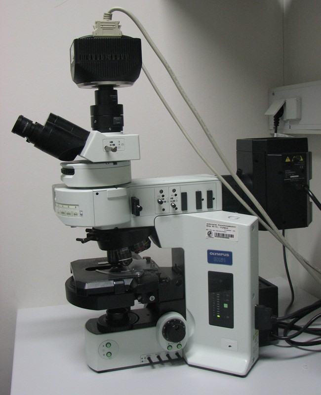
Fluorescence microscopy is a very powerful analytical tool that combines the magnifying properties of light microscopy with visualization of fluorescence. Fluorescence is a phenomenon that involves absorbance and emission of a small range of light wavelengths by a fluorescent molecule known as a fluorophore. Fluorescence microscopy is accomplished in conjunction with the basic light microscope by the addition of a powerful light source, specialized filters, and a means of fluorescently labeling a sample. This video describes the basic principles behind fluorescence microscopy including the mechanism of fluorescence, the Stoke’s shift, and photobleaching. It also gives examples of the numerous ways to fluorescently label a sample including the use of fluorescently tagged antibodies and proteins, nucleic acid fluorescent dyes with, and the addition of naturally fluorescent proteins to a specimen. The major components of the fluorescence microscope including a xenon or mercury light source, light filters, the dichroic mirror, and use of the shutter to illuminate the sample are all described. Finally, examples of some of the many applications for fluorescence microscopy are shown.


Introduction to Fluorescence Microscopy
6/3/2014
JoVE Science Education Database. General Laboratory Techniques. Introduction to Fluorescence Microscopy. Journal of Visualized Experiments, Cambridge, MA, doi: 10.3791/5040 (2014).