


Confocal Laser Endomicroscopy for in vivo Imaging
10/29/2014
Confocal Laser Endomicroscopy and Narrow-band Imaging-aided Endoscopy for in vivo Imaging of Colitis and Colon Cancer in Mice
Waldner MJ, Wirtz S, Neufert C, Becker C, Neurath MF: Confocal laser endomicroscopy and narrow-band imaging-aided endoscopy for in vivo imaging of colitis and colon cancer in mice. Nature protocols 2011, 6(9):1471-1481.
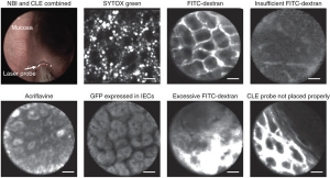
New endoscopic techniques such as narrow-band imaging (NBI) and confocal laser endomicroscopy (CLE) have improved the in vivo diagnosis of human gastrointestinal diseases in the colon. Whereas NBI may facilitate the identification of neoplastic lesions, CLE permits real-time histology of the inflamed or neoplastic colonic mucosa through the use of fluorescent dyes. These techniques have been recently adopted for use during ongoing endoscopy in mice. This protocol, which can be completed in 2 h, provides a detailed description of NBI and CLE in the mouse colon. In contrast to other techniques, this approach does not require laparotomy, and it allows direct CLE analysis of lesions identified by NBI. Mice exposed to models of colitis or colorectal cancer are anesthetized and examined with a miniaturized NBI endoscope, which provides an increased contrast of the vasculature. Upon identification of suspicious areas by NBI and the administration of fluorescent dyes, a confocal laser probe can be directed to the area of interest through the endoscope and confocal images can be obtained. Through the use of various fluorescent dyes, different aspects of the mucosa can be assessed. In addition, fluorescence-labeled antibodies can be used for molecular imaging of mice in vivo. Mouse NBI endoscopy and CLE represent reliable and fast high-quality techniques for the endoscopic characterization and molecular imaging of the mucosa in colitis and colon cancer.
 Gastrointestinal System