


T Lymphocyte Density and Distribution in Human Colorectal Mucosa, and Inefficiency of Current Cell Isolation Protocols
2/19/2016
Preza GC, Yang OO, Elliott J, Anton PA, Ochoa MT: T Lymphocyte Density and Distribution in Human Colorectal Mucosa, and Inefficiency of Current Cell Isolation Protocols. PLoS One 2015 Apr 9;10(4):e0122723.
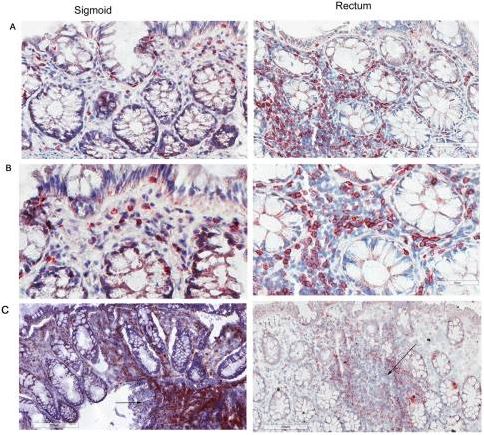
“Mucosal tissues are critical immune effector sites containing complex populations of leukocytes in a tissue microenvironment that remains incompletely understood. We identify and quantify in human distal colorectal tissue absolute mucosal CD3+ lymphocytes, including CD4+ and CD8+ subsets, by direct visualization using immunohistochemistry (IHC), immunofluorescence (IF), and an automated counting protocol (r2=0.90). Sigmoid and rectal mucosal tissues are both densely packed with T lymphocytes in the mucosal compartment. Both compartments had similar densities of CD3+ T lymphocytes with 37,400 ± 2,801 cells/mm3 and 33,700 ± 4,324 cell/mm3, respectively. Sigmoid mucosa contained 57% CD3+CD4+ and 40% CD3+CD8+ T lymphocytes which calculates to 21,300 ± 1,476/mm3 and 15,000 ± 275/mm3 T lymphocytes, respectively. Rectal mucosa had 57% CD3+CD4+ and 42% CD3+CD8+ or 21,577 ± 332, and 17,090 ± 1,206 cells/mm3, respectively. By comparison, sigmoid mucosal biopsies subjected to conventional collagenase digestion, mononuclear cell (MMC) isolation and staining for flow cytometry yielded 4,549 ± 381/mm3 and 2,708 ± 245/mm3 CD4+ and CD8+ T lymphocytes. These data suggest only ~20.7% recovery compared to IHC results for these markers. Further studies will determine if this reflects a selective bias in only CD3+, CD4+ and CD8+ T cells or can be generalized to all flow-analyzed cells from mucosal tissues for phenotyping and functional testing.“
 Gastrointestinal System