Mucosal Mononuclear Cells in Flow Cytometry Assay
6/3/2014
Quantification of Mucosal Mononuclear Cells in Tissues with a Fluorescent Bead-Based Polychromatic Flow Cytometry Assay. R. Keith Reeves, Tristan I. Evans, Jacqueline Gillis, Fay E. Wong, Michelle Connole, Angela Carville, R. Paul Johnson. Journal of Immunological Methods, Volume 367, Issues 1–2, 31 March 2011, Pages 95-98
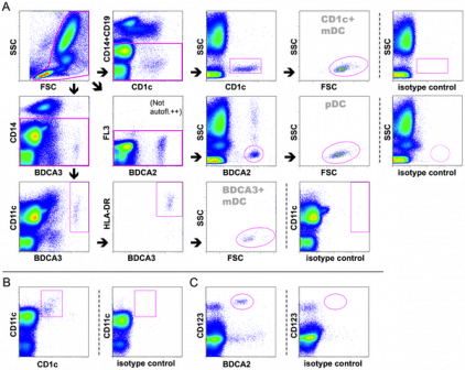
Since the vast majority of infections occur at mucosal surfaces, accurate characterization of mucosal immune cells is critically important for understanding transmission and control of infectious diseases. Standard flow cytometric analysis of cells obtained from mucosal tissues can provide valuable information on the phenotype of mucosal leukocytes and their relative abundance, but does not provide absolute cell counts of mucosal cell populations. We developed a bead-based flow cytometry assay to determine the absolute numbers of multiple mononuclear cell types in colorectal biopsies of rhesus macaques. Using 10-color flow cytometry panels and pan-fluorescent beads, cells were enumerated in biopsy specimens by adding a constant ratio of beads per mg of tissue and then calculating cell numbers/mg of tissue based on cell-to-bead ratios determined at the time of sample acquisition. Testing in duplicate specimens showed the assay to be highly reproducible (Spearman R=0.9476, P<0.0001). Using this assay, we report enumeration of total CD45(+) leukocytes, CD4(+) and CD8(+) T cells, B cells, NK cells, CD14(+) monocytes, and myeloid and plasmacytoid dendritic cells in colorectal biopsies, with cell numbers in normal rhesus macaques varying from medians of 4486 cells/mg (T cells) to 3 cells/mg (plasmacytoid dendritic cells). This assay represents a significant advancement in rapid, accurate quantification of mononuclear cell populations in mucosal tissues and could be applied to provide absolute counts of a variety of different cell populations in diverse tissues.


