Histological Sample Preparation for Light Microscopy
6/3/2014
JoVE Science Education Database. General Laboratory Techniques. Histological Sample Preparation for Light Microscopy. Journal of Visualized Experiments, Cambridge, MA, doi: 10.3791/5039 (2014).
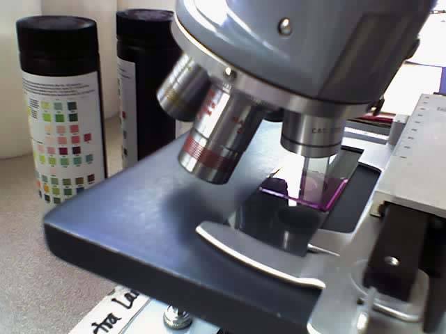
Histology is the study of cells and tissues, which is typically aided by the use of a light microscope. The preparation of histological samples can vary greatly based on the inherent properties of the samples such as size and hardness as well as expected post-processing which includes planned staining techniques or other down-stream applications. As described in this video, specimen preparation typically begins with a fixation procedure to prevent degradation of the sample by naturally occurring enzymes that are released by the cells upon death. Once fixed, samples are placed into an embedding medium that is able to sufficiently support the sample. Most commonly this is paraffin wax, but other materials such as a glycerin based freezing medium and agars are also used to surround the sample during sectioning. Sectioning then takes place on a microtome or other cutting device that allows the user to shave the sample into thin slices ranging from a few microns to a few millimeters in thickness. Once cut, sections are mounted on a glass slide and stained to bring out specific features of the sample before being imaged on a microscope.