


Formation of Human Colonic Crypt Array
3/9/2018
Formation of Human Colonic Crypt Array by Application of Chemical Gradients Across a Shaped Epithelial Monolayer. Wang, Yuli et al. Cellular and Molecular Gastroenterology and Hepatology, Volume 5 , Issue 2 , 113 – 130. DOI: https://doi.org/10.1016/j.jcmgh.2017.10.007.
In this study, Wang, Allbritton and colleagues have used human colonic organoids to generate physiologically relevant “organ on a chip” cultures for high throughput analyses. By growing the epithelial cells on a structured hydrogel scaffold and applying growth factors Wnt-3A, R-spondin, and noggin to the basal media only, they achieved compartmentalization of the stem/progenitor cells, appropriate lineage differentiation and cell movement that closely replicated human crypt biology. This new system offers the easy accessibility of monolayer cultures while retaining the 3-dimensional architecture and tissue polarity of in vivo colon crypts.
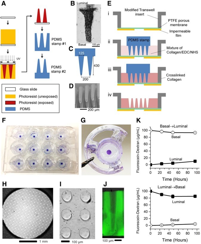
Pictured: Scaffolding and gradient cassette for the culture of in vitro crypts. (A) Fabrication process for the PDMS stamps used to micromold collagen scaffolds mimicking the topography of epithelium of human colon. (B) Transmitted light microscopy of a human colonic crypt from a biopsy sample. (C) Geometry of the PDMS stamp that was used to micromold collagen. Units are shown in micrometers. (D) SEM image of the PDMS stamp. (E) Schematic of the micromolding process used to generate a shaped, cross-linked collagen scaffold on a modified Transwell insert. (F) Image of the 12 scaffolds within a 12-well plate. To show the trans-scaffold transport occurred only at the central area, the insert was briefly exposed to 0.1% toluidine blue from the basal side. The toluidine blue only stained the scaffold within the central 3-mm diameter area in contact with the basal compartment. (G) Image of a single scaffold within an insert. (H) Top view of an array of microwells created in the scaffold. (I) Close-up view of a region of the scaffold in panel H. (J) Side view of a cross-section through a fluorescein-labeled collagen scaffold. (K) Concentration of fluorescein dextran (molecular weight, 40 kilodaltons) measured at the luminal (solid squares) and basal (empty circles) compartments at 24, 48, and 72 hours. Top plot: Diffusion from basal to luminal. At 0 hours, 0.5 mL PBS was added to the luminal compartment and 1.5 mL fluorescein dextran (100 μg/mL) was added to the basal compartment. Bottom plot: Diffusion from luminal to basal. At 0 h, 0.5 mL fluorescein dextran (100 μg/mL) was added to the luminal compartment and 0.5 mL PBS was added to the basal compartment. Cellular and Molecular Gastroenterology and Hepatology 2018 5, 113-130DOI: (10.1016/j.jcmgh.2017.10.007).
 Gastrointestinal System